
Abnormal Thermology
Talar Dome Osteochondritis With Peroneal Tendinosis and a Spinal Cord Stimulator
Dr. Robert Schwartz
Posted on January 30, 2018, updated on September 30, 2019
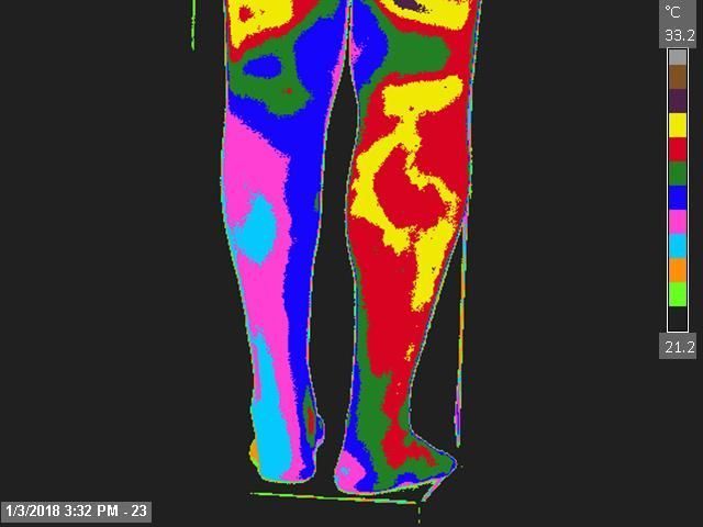
29 yo with a CC of LLE pain after rolling his ankle. MRI revealed talar dome osteochondritis, peroneus brevis and Longus tendon tear and tendinosis, with lateral malleolus contusion. Patient underwent ankle arthroscopy. Post-operative MRI revealed persistent talar dome osteochondral defect. A spinal cord stimulator was placed at L4 & S1 for RSD. Cold stress Thermography revealed localized asymmetry over the posterior, lateral and anterior leg, the posterior and lateral foot/ankle, and a isolated spot over L2. Thermographic Impression was a cold LLE distal to the knee in the presence of an isolated hot spot over the L2 spinal level.
Supporting Documents