
Abnormal Thermology
Computer Aided Thermography to Detect Malignancy of the Breast
Geetha Manjunath
Published on August 1, 2022
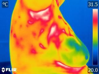
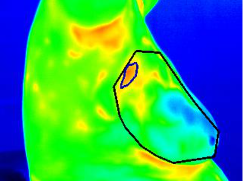
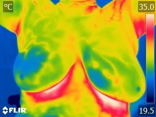
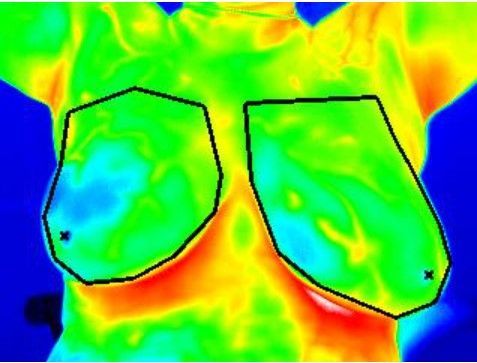
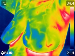
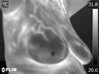
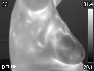
A 64-year-old post menopausal female with pain in her right breast, with first degree relative father had liver cancer. The expert thermologist graded the heat pattern as normal despite the abnormal heat pattern near the axillary tail. However when the same images were presented along with SMILE-100 annotations, which clearly showed the thermal increase as a Hotspot, the thermologist suspected malignancy and suggested a diagnostic workup for the participant. An irregular hypoechoic lesion was found in the right breast at the 10 o’clock position after breast ultrasonography investigation. Lumpectomy of the right breast confirmed the lesion as invasive ductal carcinoma (Grade 2) of no special type.

Supporting Documents
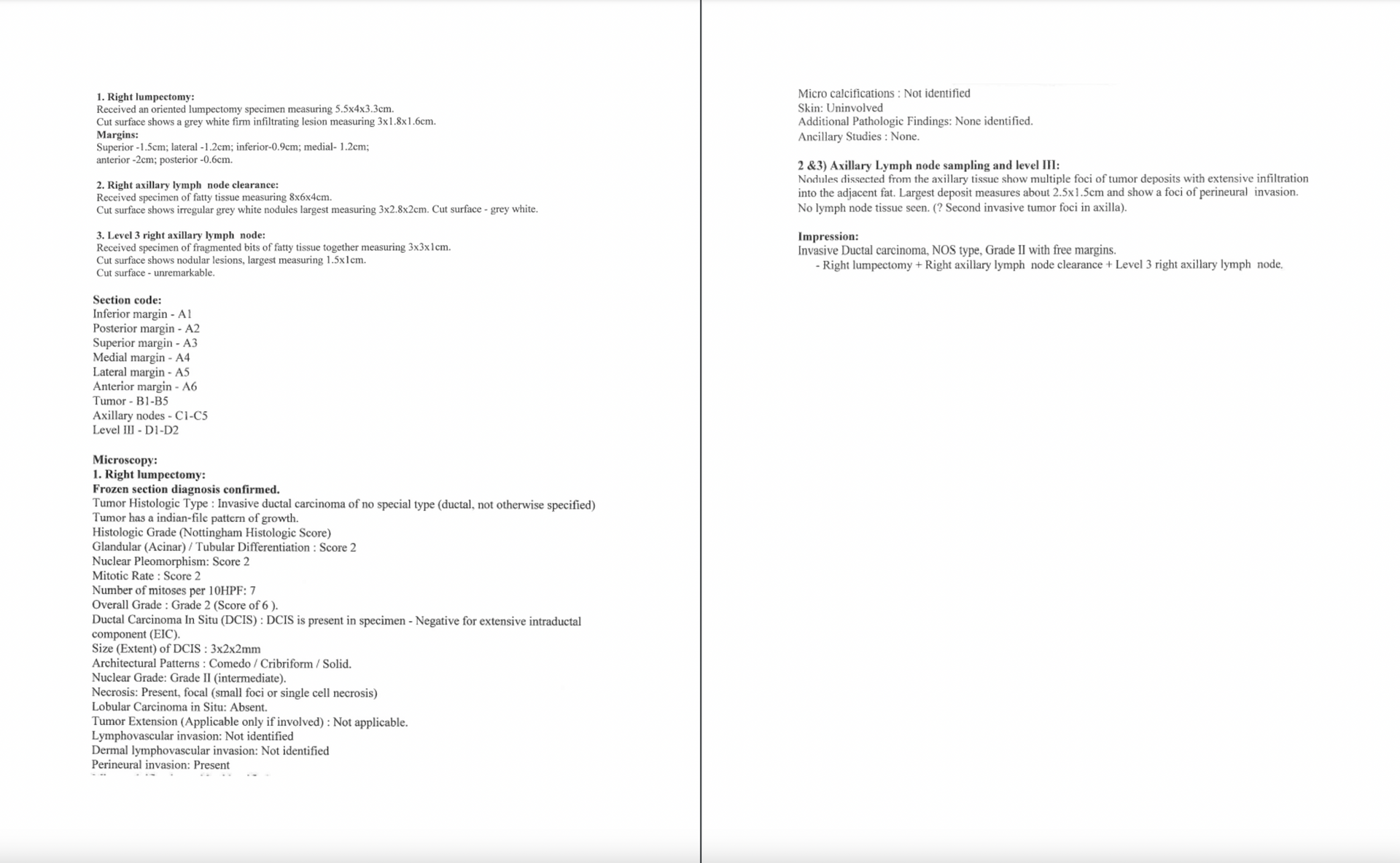
Histopathology
Abnormal